|
|
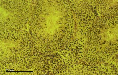
Testis, mouse - histology slide
|
Testicles: Cross-section; mouse. Optical microscopy technique: Bright field. Magnification: 600x (for picture width 26 cm ~ A4 format).
Histology slide courtesy of Wikimedia Commons and Doc. RNDr. Josef Reischig, CSc.
This file is licensed under the Creative Commons Attribution-Share Alike 3.0 Unported license.
|
|