|
|
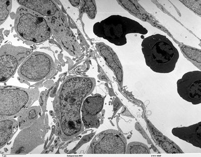
Embryonic brain 80437
|
Transmission electron microscope image of a thin section cut through the developing brain tissue (telencephalic hemisphere) of an 11.5 day mouse embryo. This image shows the telencephalon surface just before perforation by a capillary. Before perforation occurs, the vascular and cerebral basal laminae are in contact and will become fused at the point of perforation by the the capillary filopodia. JEOL 100CX TEM
Image courtesy of Louisa Howard, Miguel Marin-Padilla.
|
|