|
Histology-World!
|
|
|
|
|
Key Histology FeaturesInstructions: Run your mouse over the histology slide want to view. Key histology features are described. If a link is present, click to view and listen to the histology audioslide.
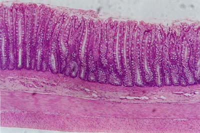
Large IntestineOn histology slides (1), (2) and (3),
three layers of the large intestine are visible: mucosa,
submucosa and muscularis externa. On histology slide (1) and (2), the two distinct layers of
muscle on the muscularis externa are visible: the inner circular and
an outer longitudinal. Notice that there are no villi
here, in contrast to the small intestine. The apical surface is smooth, without
finger like projections. There are lots of goblet cells on the mucosa.
These are the clear looking hollowed out spaces.
|
| Copyright (c) Histology-World and its licensors. All rights reserved. |