|
|
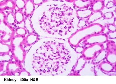
Kidney - histology slide
|
This histology image shows two glomeruli and adjacent tubules. Each glomerulus is surrounded by Bowman's capsule. The glomerular tuft consists of a network of endothelially lined capillaries with a thin basement membrane. Occasional small clusters of cells surrounded by eosinophilic material represent the mesangium which supports the capillaries. The tubules, mainly proximal convoluted, are lined by a single layer of cuboidal or low columnar epithelium.
Histology image descriptions and photography by Frank N. Miller, M.D.; histology image courtesy of Uniformed Services University of the Health Sciences.
|
|