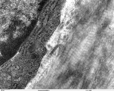
Histology-World!
Histology Photo Album
Home
Login
Histology Photo Album Home
Album list
Last uploads
Last comments
Most viewed
Top rated
My Favorites
Search
FILE 14/115
Kneecap - TEM
Transmission electron microcope image of a thin section cut through the cartilage/bone interface in a mouse kneecap. Image shows striated type I collagen fibers and the edge of an osteoblast cell.
JEOL 100CX TEM
Image courtesy of Louisa Howard and Roy Fava.
File information
Filename:
kneecap_interface_80005-33.jpg
Album name:
sarah
/
Bone Histology
Keywords:
EM
Filesize:
676 KiB
Date added:
Aug 04, 2007
Dimensions:
1560 x 1246 pixels
Displayed:
5007 times
URL:
http://histology-world.com/photoalbum/displayimage.php?pid=1012
Favorites:
Add to Favorites
Copyright Histology-World and its licensors. All rights reserved
.