|
|
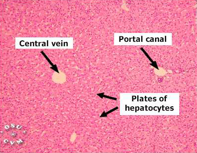
Liver (labels) - histology slide
|
This is a histology slide of the liver.
Sinusoids are larger than conventional capillaries and less regular in shape. They
are lined by thin endothelial cells (E). Also residing on the sinusoidal walls are
macrophages called Kupffer cells (K). These are phagocytic cells that remove
particulate material and old red blood cells from circulation. Kupffer cells are
members of the mononuclear phagocyte system.
Histology image courtesy of OSU College of Veterinary Medicine. Copyright © Charlotte L. Ownby. Reprinted with permission.
|
|