|
|
Lung (labels) - histology illustration
|
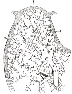
Part of a secondary lobule from the depth of a human lung, showing parts of several primary lobules.
1, bronchiole; 2, respiratory bronchiole; 3, alveolar duct; 4, atria; 5, alveolar sac; 6, alveolus or air cell: m, smooth muscle; a, branch pulmonary artery; v, branch pulmonary vein; s, septum between secondary lobules. Camera drawing of one 50 μ section. X 20 diameters. (Miller.)
Histology illustration courtesy of Gray's Anatomy.
|
|