|
|
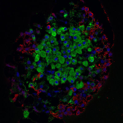
Islet of Langerhans - laser scanning confocal microscope image
|
Islet of Langerhans isolated from rat pancreas. Laser scanning confocal microscope image. 63x, oil imm. objective.
Colors explanation:
* Nuclei stained blue with DAPI
* Insuline (beta-cells) stained green with anti-insuline dye conjugated Abs
* Glucagon (alpha-cells) stained red with anti-glucagon dye conjugated Abs
Image courtesy of Masur. Permission is granted to copy, distribute and/or modify this document under the terms of the GNU Free Documentation License, Version 1.2 or any later version published by the Free Software Foundation; with no Invariant Sections, no Front-Cover Texts, and no Back-Cover Texts. A copy of the license is included in the section entitled "GNU Free Documentation License".
|
|