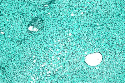
Micrograph of the liver showing a portal tract and a nearby hepatic venule. Reticulin stain. The normal "hepatocyte plate" -- the strands of hepatocytes that characterise the morphology of the liver and are separated by the liver sinusoids -- is one or two cells thick, as shown in the image. (Description text from the Wikimedia Commons file page.)