|
|
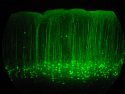
Somatosensory cortex
|
20X magnification of the somatosensory cortex of a mouse brain slice. These little guys have green flourescent protein (GFP) from a jelly-fish expressed in a subset of their neurons. Layer V neuron cell bodies are the teardrop shaped things at the bottom and then the dendrite reaches up like a tree to then bifurcate near the top (pial surface).
Attribution-Share Alike 2.0 Generic
You are free:
· to Share - to copy, distribute, display, and perform the work
· to Remix - to make derivative works
Under the following conditions:
· Attribution. You must attribute the work in the manner specified by the
author or licensor (but not in any way that suggests that they endorse you
or your use of the work).
· Share Alike. If you alter, transform, or build upon this work, you may
distribute the resulting work only under the same or similar license to this
one.
· For any reuse or distribution, you must make clear to others the license
terms of this work. The best way to do this is with a link to this web page:
http://creativecommons.org/licenses/by-sa/2.0/deed.en-us.
· Any of the above conditions can be waived if you get permission from the
copyright holder.
· Nothing in this license impairs or restricts the author's moral rights.
|
|