|
|
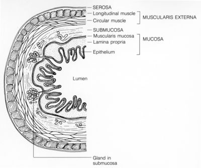
GI (labels) - histology illustration
|
Line drawing showing the lining of the GI tract (colorectal). The walls of the digestive tract have four layers of tissue: mucosa, submucosa, muscularis externa and serosa.
Histology illustration courtesy of National Cancer Institute.
|
|