|
|
Cornea (labels) - histology illustration
|
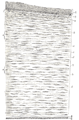
Vertical section of human cornea from near the margin. (Waldeyer.) Magnified. 1. Epithelium. 2. Anterior elastic lamina. 3. substantia propria. 4. Posterior elastic lamina. 5. Endothelium of the anterior chamber. a. Oblique fibers in the anterior layer of the substantia propria. b. Lamellæ the fibers of which are cut across, producing a dotted appearance. c. Corneal corpuscles appearing fusiform in section. d. Lamellæ the fibers of which are cut longitudinally. e. Transition to the sclera, with more distinct fibrillation, and surmounted by a thicker epithelium. f. Small bloodvessels cut across near the margin of the cornea.
Histology illustration courtesy of Gray's Anatomy.
|
|