|
|
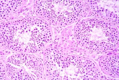
Testes - histology slide
|
This is a histology slide of the testis. The seminiferous tubules are arranged in lobules separated by fibrous septa. In the interstitium between tubules Leydig cells with eosinophilic cytoplasm are seen. Each tubule has a thin fibrous wall. Tubules contain germ cells which mature from the outer portion toward the center, where there are numerous spermatozoa. Sertoli cells are characterized by a pale nucleus and a nucleolus.
Histology slide descriptions and photography by Frank N. Miller, M.D.; histology slide courtesy of Uniformed Services University of the Health Sciences.
|
|