|
|
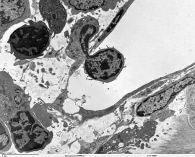
Kneecap - TEM
|
Transmission electron microscope image of a thin section cut through the cartilage/bone interface in a mouse kneecap. Blood cells are located in the upper part of image, just adjacent to the bone/cartilage interface. JEOL 100CX TEM
Image courtesy of Louisa Howard and Roy Fava.
|
|